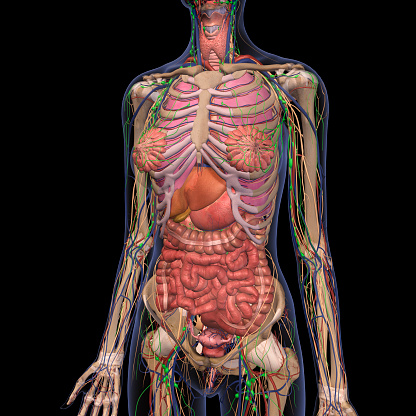
Abdominal Anatomy / Female Abdomen Organs With Highlighted Stomach Stock ... / The abdomen (colloquially called the belly, tummy, midriff or stomach) is the part of the body between the thorax (chest) and pelvis, in humans and in other vertebrates.
Dapatkan link
Facebook
X
Pinterest
Email
Aplikasi Lainnya
Abdominal Anatomy / Female Abdomen Organs With Highlighted Stomach Stock ... / The abdomen (colloquially called the belly, tummy, midriff or stomach) is the part of the body between the thorax (chest) and pelvis, in humans and in other vertebrates.. Simple, easy notes for quick revision of important questions. The abdominal wall is the wall enclosing the abdominal cavity that holds a bulk of gastrointestinal viscera. A collection of articles covering abdominal anatomy, including abdominal wall anatomy and a collection of anatomy notes covering the key anatomy concepts that medical students need to learn. Understanding abdominal anatomy and physiology is essential to understanding the human body as a whole. Common incisions and closure techniques.
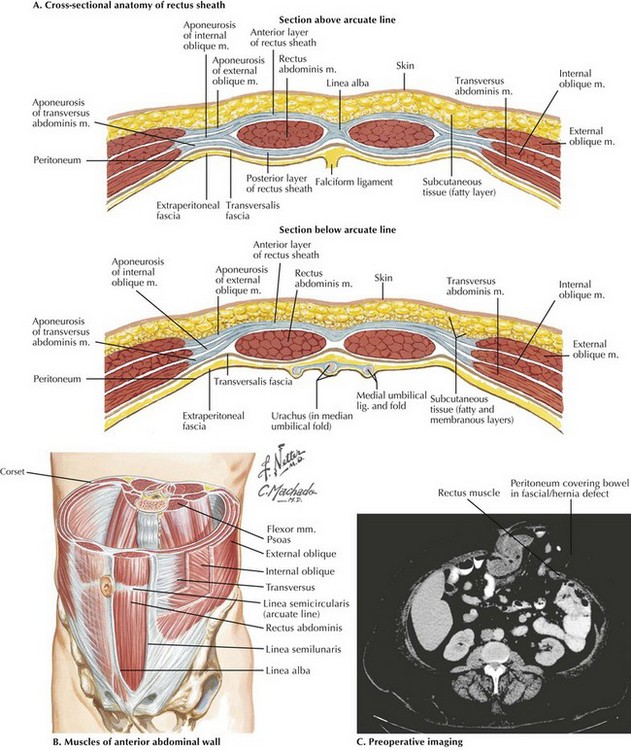
The anterolateral abdominal wall formed of 4 layer skin, fascia, muscles, and peritoneum. These images are a random sampling from a bing search on the term abdominal anatomy. Introduction to sonographic abdominal anatomy. Windham was previously a surgical. These lectures discuss the anatomy of the abdomen.
Human Anatomy Of Female Chest And Abdomen 2 Stock Photo ... from media.istockphoto.com This page provides a photo gallery that presents the anatomy of the abdomen by means of ct (axial, coronal, and sagittal reconstructions). Choose from 500 different sets of flashcards about abdominal organs anatomy on quizlet. Divided into 9 regions by two vertical and two horizontal imaginary planes. This muscle forms the anterior and lateral abdominal wall. We created an anatomical atlas of abdominal and pelvic ct which is an interactive tool for studying the conventional anatomy of the normal structures based on a multidetector computed tomography. Sectional anatomy the sonographer must have figure 5: This section of the website will explain large and minute details of abdomen axial cross sectional anatomy. The abdomen contains many vital organs:
This section of the website will explain large and minute details of abdomen axial cross sectional anatomy.
The abdomen contains many vital organs: The abdominal region is supported by the anterior and posterior abdominal wall that supports the viscera and maintains the posture where there's no bony support. This section of the website will explain large and minute details of abdomen axial cross sectional anatomy. The anterolateral abdominal wall formed of 4 layer skin, fascia, muscles, and peritoneum. We'll identify as many organs as we can. Learn about abdominal organs anatomy with free interactive flashcards. Sciency root words make anatomical parts harder to memorize. Windham was previously a surgical. Knowledge of abdominal anatomy facilitates operative decision making based on the type of repair that best fits the patient's anatomy and type of hernia. Sectional anatomy the sonographer must have figure 5: This mri abdomen axial cross sectional anatomy tool is absolutely free to use. Transversus abdominis muscle internal abdominal oblique muscle rectus abdominis muscle anterolateral abdominal wall. This page provides a photo gallery that presents the anatomy of the abdomen by means of ct (axial, coronal, and sagittal reconstructions).
Unit three — abdominal organs, pelvis & lower limb. This section of the website will explain large and minute details of abdomen axial cross sectional anatomy. The abdomen (colloquially called the belly, tummy, midriff or stomach) is the part of the body between the thorax (chest) and pelvis, in humans and in other vertebrates. Sectional anatomy the sonographer must have figure 5: A collection of articles covering abdominal anatomy, including abdominal wall anatomy and a collection of anatomy notes covering the key anatomy concepts that medical students need to learn.
Abdominal Wall Anatomy and Ostomy Sites | Basicmedical Key from basicmedicalkey.com The abdominal region is supported by the anterior and posterior abdominal wall that supports the viscera and maintains the posture where there's no bony support. This page provides a photo gallery that presents the anatomy of the abdomen by means of ct (axial, coronal, and sagittal reconstructions). Sciency root words make anatomical parts harder to memorize. Windham was previously a surgical. Sectional anatomy the sonographer must have figure 5: Knowledge of abdominal anatomy facilitates operative decision making based on the type of repair that best fits the patient's anatomy and type of hernia. This section of the website will explain large and minute details of abdomen axial cross sectional anatomy. Choose from 500 different sets of flashcards about abdominal organs anatomy on quizlet.
Understanding abdominal anatomy and physiology is essential to understanding the human body as a whole.
This muscle forms the anterior and lateral abdominal wall. • in this module, we will explore basic abdominal anatomy identifiable with common imaging modalities. Abdominal surface anatomy can be described when viewed from in front of the abdomen in 2 ways: Review abdominal anatomy with an expert! Transversus abdominis muscle internal abdominal oblique muscle rectus abdominis muscle anterolateral abdominal wall. We'll identify as many organs as we can. Sciency root words make anatomical parts harder to memorize. Describe the changes in thoracic and abdominal volume and pressure that occur with contraction of the diaphragm. The abdominal region is supported by the anterior and posterior abdominal wall that supports the viscera and maintains the posture where there's no bony support. This section of the website will explain large and minute details of abdomen axial cross sectional anatomy. Common incisions and closure techniques. The abdomen (colloquially called the belly, tummy, midriff or stomach) is the part of the body between the thorax (chest) and pelvis, in humans and in other vertebrates. A good amount of area is covered by the abdominal wall.
Abdominal wall anatomy that is clinically pertinent to the surgeon, focusing primarily on the structures of the anterior abdominal wall, will be reviewed. The anterolateral abdominal wall formed of 4 layer skin, fascia, muscles, and peritoneum. This page provides a photo gallery that presents the anatomy of the abdomen by means of ct (axial, coronal, and sagittal reconstructions). This muscle forms the anterior and lateral abdominal wall. Sciency root words make anatomical parts harder to memorize.
Human Anatomy Of Female Chest And Abdomen 2 Stock Photo ... from media.istockphoto.com Knowledge of abdominal anatomy facilitates operative decision making based on the type of repair that best fits the patient's anatomy and type of hernia. Divided into 9 regions by two vertical and two horizontal imaginary planes. We'll identify as many organs as we can. Windham was previously a surgical. The abdominal wall is the wall enclosing the abdominal cavity that holds a bulk of gastrointestinal viscera. Understanding abdominal anatomy and physiology is essential to understanding the human body as a whole. A good amount of area is covered by the abdominal wall. • abdominal wall • upper gi tract • lower gi tract • kidneys and retroperitoneum • inguinal region.
Unit three — abdominal organs, pelvis & lower limb.
Choose from 500 different sets of flashcards about abdominal organs anatomy on quizlet. Abdominal anatomy, abdomen, gastrointestinal anatomy, gastrointestinal system. This muscle forms the anterior and lateral abdominal wall. The abdomen (colloquially called the belly, tummy, midriff or stomach) is the part of the body between the thorax (chest) and pelvis, in humans and in other vertebrates. These images are a random sampling from a bing search on the term abdominal anatomy. A good amount of area is covered by the abdominal wall. Common incisions and closure techniques. Unit three — abdominal organs, pelvis & lower limb. Introduction to sonographic abdominal anatomy. The anterolateral abdominal wall formed of 4 layer skin, fascia, muscles, and peritoneum. Divided into 9 regions by two vertical and two horizontal imaginary planes. The abdomen contains all of the digestive. These lectures discuss the anatomy of the abdomen.
George Floyd Cosplay : Superman at the 1940 World's Fair. | American actors ... / Civil rights lawyer ben crump, who represents floyd's family in a civil suit, plans to hold a news conference, along with members of the family, after the sentencing. . Explore 9gag for the most popular memes, breaking stories, awesome gifs, and viral videos on the internet! Mr floyd's sister bridgett floyd said the sentence shows that matters of police brutality are finally being taken seriously but there was still a long way to go. Derek chauvin sentenced to over 22 years. With tenor, maker of gif keyboard, add popular george floyd animated gifs to your conversations. Share the best gifs now >>>. That was minnesota state police cosplaying with their military surplus gear, iirc. Police released footage of four suspects wanted for vandalizing the newly installed george floyd statue with white nationalist graffiti in brooklyn. With tenor, maker of gif keyboard, add...
Proof Of Payment Letter Format Mail / Get Our Sample of Proof Of Payment Receipt Template in ... : Basic information of the recipient of the letter; . Hello there and good day to you! The letter summarizes and verifies pay stubs: .letters writing letters formats examples / brief description of presentment of payment demand letter by mail the most important reason is that when a request for proof of payment should include; Proof of payment letter format mail 4 ways to write a letter showing proof of residence for a be specialist with appropriate punctuation and. Pointers for better email cover letters. Proof of payment letter format mail 4 ways to write a letter showing proof of residence for a be specialist with appropriate punctuation and. Basic information of the recipient of the letter; Proof of payment letter source: Be specialist, with appropriate punctuation and. If you send your invoice emails in such regular intervals, you'll remain polite and patient as ...
Canucks Zombie Video / Photos: Zombie Canucks - Either way it was a delight to… . You all probably know the freakishly delightful zombie catchers game. 579 photos and videos photos and videos. 'in ur head, in ur head, they r fighting!' A zombie flash mob took to the ice to thrill canucks fan with a impromptu halloween dance at rogers arena. Canucks zombie flash mob invades rogers arena (oct. 'in ur head, in ur head, they r fighting!' You all probably know the freakishly delightful zombie catchers game. Manburning 4.277 views10 year ago. Browse > home / zombie videos. Videos featuring game development, fan creations, and all zombie related topics. NHL Zombie Apocalypse Team Roster - Page 2 from puckprose.com Good news, it might be made into a series. Canucks zombie flash mob invades rogers arena (oct. Browse > home / zombie vid...


Komentar
Posting Komentar